Immunohistochemistry (IHC)/ Immunocytochemistry (ICC)
Immunohistochemistry (IHC)/ Immunocytochemistry (ICC) are the most common applications of immunostaining. IHC is concerned with labeling tissue, whereas ICC is used to identify specific proteins or antigens in cells.
The principle for both techniques is the same, identifying unique target antigens with labeled antibodies that specifically bind the desired target.
There are direct methods (one-step staining methods) or indirect methods of detection. The former has a tagged antibody, usually a fluorescent molecule (e.g. FITC Cat. No. 5440) or chromagen/substrate (e.g. horseradish peroxidase (HRP)), which directly binds an antigen of interest. The latter involves primary antibodies binding the target antigen, and a labeled secondary antibody binding the primary antibody.
The direct method is rapid and simple but is much less sensitive than the indirect method. The indirect method is particularly useful for detecting a target that is expressed at low levels.
Immunohistochemistry (IHC)/ Immunocytochemistry (ICC) principles: Direct & Indirect Methods

| Steps | Direct Method | Indirect Method |
|---|---|---|
| 1 | Label antigen with specific primary antibody | Label antigen with specific primary antibody |
| 2 | Wash excess primary antibodies off | Wash excess primary antibodies off |
| 3 | Visualize sample on e.g. fluorescence microscope | Label with tagged secondary antibody |
| 4 | - | Wash excess secondary antibodies off |
| 5 | - | Visualise sample on i.e. fluorescence microscope |
Tyramide Signaling Amplification (TSA) in IHC & ICC
Tyramide Signal Amplification (TSA), also known as Catalyzed Reported Deposition (CARD), efficiently amplifies signal and detection capabilities for low-abundance targets in ICC and IHC applications.
Horseradish peroxide (HRP) catalyzes the transformation of labeled inactive tyramides (e.g. Biotinyl Tyramide Cat. No. 6241) into reactive radicals, which then bind to adjacent tyrosine residues significantly increasing the density of labeling. Tyramide reagents can be conjugated with a fluorescent dye or a hapten depending on detection methods used.

Figure 1: Tyramide Signal Amplification Principles: Tissue or cell samples are labeled with primary and secondary antibodies. The secondary antibody, which is pre-conjugated to horseradish peroxidase (HRP), is exposed to H2O2. This catalyzes the transformation of a labeled tyramide substrate into a highly reactive species that binds to tyrosine residues on the proteins in close proximity to the antibodies and HRP, amplifying the signal.
IHC and ICC Counterstains
After the initial labeling step in ICC and IHC, a second stain termed a counterstain is often applied, which helps provide contrast so that the primary label can be seen more clearly. Depending on the experimental requirements, counterstains can either show high specificity for specific types of biomolecules, such as DNA or cytoskeleton, or stain the whole cell. Hematoxylin ( Cat. No. 5222), Hoechst stain (Hoechst 33342 Cat. No. 5117),and DAPI (Cat. No. 5748) are commonly used counterstains. It is important that when choosing counterstains, they have minimum spectral overlap with the fluorochrome conjugated to the detection antibody.
IHC and ICC Blocking Buffers
To reduce non-specific binding of primary or secondary antibodies, blocking buffers made with reagents such as Bovine Serum Albumin (BSA) (Cat. No. 5217) are applied to block non-target binding sites. In addition to using blocking buffers, incubation times and temperatures can also be varied to maximize specific binding and thus the optimum detection window for any antibody set.
Selecting Dyes for Multicolor Immunostaining
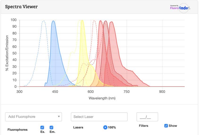
When selecting multiple fluorescent dyes for immunostaining, it is important to have the smallest spectral overlap to give the clearest results. Use our spectra viewer to plan your experiment.

Bio-Techne Antibodies for Immunofluorescence Assays
Bio-Techne brands R&D Systems™ and Novus Biologicals provide the highest quality antibodies giving you repeatedly reliable results. Our comprehensive portfolio comprises over 40,000 IHC validated antibodies covering the most common and unique targets. Our products are highly cited in peer-reviewed journals.
Bio-Techne's Immunohistochemistry Handbook
This resource gives a comprehensive introduction to IHC that is well-suited for researchers new to the technique or experienced scientists looking for the latest innovations or a quick refresher. Download or request a copy.

Literature for Immunohistochemistry (IHC)/ Immunocytochemistry (ICC)
Tocris offers the following scientific literature for Immunohistochemistry (IHC)/ Immunocytochemistry (ICC) to showcase our products. We invite you to request* your copy today!
*Please note that Tocris will only send literature to established scientific business / institute addresses.
Fluorescent Dyes and Probes Research Product GuideUpdated
This product guide provides a background to the use of Fluorescent Dyes and Probes, as well as a comprehensive list of our:
- Fluorescent Dyes, including Janelia Fluor® Dyes
- Fluorescent Probes for Advanced Microscopy and Live Cell Imaging
- Fluorescent Probes to Support In Vivo, Deep Tissue, and Bioluminescence Imaging
- Fluorescent Probes and Reagents for Organoids and 3D Cell Culture Imaging
- Aptamer-based RNA Imaging Reagents
- Fluorescent Probes for Imaging Bacteria
- TSA VividTM Fluorophore Kits
- TSA Reagents for Enhancing IHC, ICC & FISH Signals
- Tissue Clearing Kits and Reagents
