Submit a Review & Earn an Amazon Gift Card
You can now submit reviews for your favorite Tocris products. Your review will help other researchers decide on the best products for their research. Why not submit a review today?!
Submit ReviewKey information: Phalloidin-Janelia Fluor® 646 is a red fluorescent F-actin probe. It binds and labels specifically F-actin.
Used for: Phalloidin-Janelia Fluor® 646 can be used to visualize and quantify F-actin in tissue sections, cell cultures or cell-free experiments with optimal results in formaldehyde-fixed and permeabilized cells.
Application: confocal microscopy, super-resolution microscopy (SRM) including dSTORM and STED and flow cytometry.
Properties and Photophysical Data: Phalloidin-Janelia Fluor® 646 is composed of the F-actin probe, Phalloidin (Cat. No. 4535), conjugated to Janelia Fluor® 646 (Cat. No. 6148). Excitation and emission maxima (λ) are 646 nm and 664 nm, respectively; quantum yield = 0.54; extinction coefficient = 152,000 M-1cm-1.
 View Larger
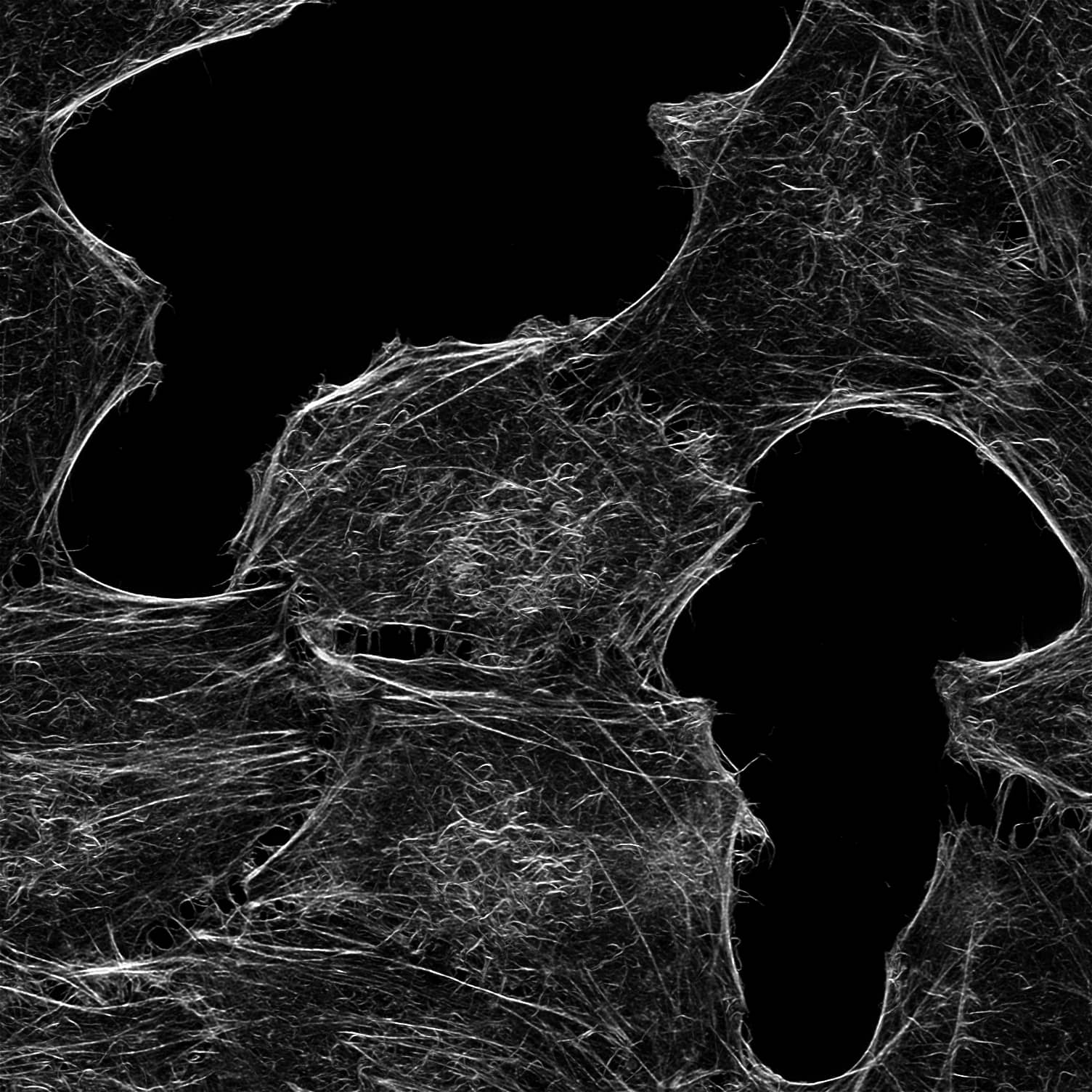
Application of Phalloidin-Janelia Fluor® 646. HeLa cells were fixed for 10 minutes with 4% formaldehyde in PBS and permeabilized with PBS/0.25% Triton X-100 for 6 minutes. An additional post-fixation step was performed twice with 1% formaldehyde in PBS followed by 3 washes with PBS. Phalloidin Janelia Fluor® 646 was prepared as a 10 μM stock solution diluted in methanol and diluted to a working concentration of 20 nM in PBS. Stained cells were imaged with a Leica SP8X WLL upright confocal microscope. Image kindly provided by Brigitte Bergner, Steffen Dietzel, Core Facility Bioimaging at the Biomedical Center of the Ludwig-Maximilians-Universität München.
Sold under license from the Howard Hughes Medical Institute, Janelia Research Campus
Use our spectra viewer to interactively plan your experiments, assessing multiplexing options. View the excitation and emission spectra for our fluorescent dye range and other commonly used dyes.
Spectral Viewer| M. Wt | 1266.51 |
| Formula | C64H75N11O13SSi |
| Storage | Store at -20°C |
| Purity | ≥90% (HPLC) |
| InChI Key | AOXMEZGPCIYGOB-BNAQDRCBSA-N |
| Smiles | O=C([C@H](NC([C@H](C)NC([C@H](C[C@](O)(C)CNC(C1=CC=C(C([O-])=O)C(C2=C(C=C/3)C([Si](C)(C)C4=CC(N5CCC5)=CC=C42)=CC3=[N+]6CCC\6)=C1)=O)N7)=O)=O)[C@H](C)O)N[C@H]8CSC(N9)=C(C[C@H](NC([C@H](C)NC([C@@H]%10C[C@@H](O)CN%10C8=O)=O)=O)C7=O)C%11=C9C=CC=C%11 |
The technical data provided above is for guidance only. For batch specific data refer to the Certificate of Analysis.
Tocris products are intended for laboratory research use only, unless stated otherwise.
References are publications that support the biological activity of the product.
Grimm et al (2015) A general method to improve fluorophores for live-cell and single-molecule microscopy. Nat.Methods 12 244 PMID: 25599551
Cooper et al (1987) Effects of cytochalasin and phalloidin on actin. J.Cell.Biol. 105 1473 PMID: 3312229
Keywords: Phalloidin-Janelia Fluor 646, Phalloidin-Janelia Fluor 646 supplier, Phalloidin, Janelia, Fluor, 646, red, fluorescent, F-actin, probes, dyes, stains, cytoskeletal, cytoskeleton, Phalloidin-JF646, Fluorescent, Actin, Probes, 7201, Tocris Bioscience
Citations are publications that use Tocris products.
Currently there are no citations for Phalloidin-Janelia Fluor® 646.
There are currently no reviews for this product. Be the first to review Phalloidin-Janelia Fluor® 646 and earn rewards!
$25/€18/£15/$25CAN/¥75 Yuan/¥2500 Yen for a review with an image
$10/€7/£6/$10 CAD/¥70 Yuan/¥1110 Yen for a review without an image
Tocris offers the following scientific literature in this area to showcase our products. We invite you to request* your copy today!
*Please note that Tocris will only send literature to established scientific business / institute addresses.
This product guide provides a background to the use of Fluorescent Dyes and Probes, as well as a comprehensive list of our: