Submit a Review & Earn an Amazon Gift Card
You can now submit reviews for your favorite Tocris products. Your review will help other researchers decide on the best products for their research. Why not submit a review today?!
Submit ReviewRNA Imaging Probe 1c is a fluorogenic RNA imaging probe. RNA Imaging Probe 1c displays high membrane permeability, strong fluorogenic responses upon binding RNA, compatibility with fluorescence lifetime imaging microscopy (FLIM), low cytotoxicity, and excellent photostability. Excitation and emission maxima (λ) are 556 nm and 608 nm, respectively; quantum yield = 0.49; extinction coefficient = 27,500 M-1cm-1. Suitable for live cell imaging.
 View Larger
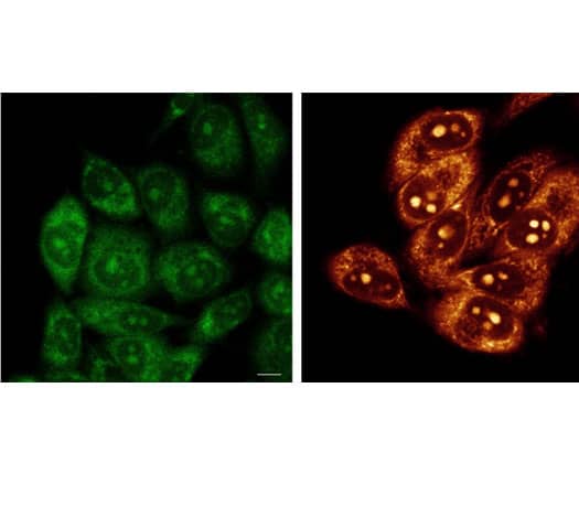
Application of RNA Imaging Probe 1c on live HeLa cells. Confocal fluorescence images of live Hela cells imaged with 20 μM methyl pyridinium indole (MPI) (green) ( lambda ex = 470 nm, lambda em = 525-555 nm) and 20 μM RNA Imaging Probe 1c (red) ( lambda ex = 550 nm, lambda em= 580-620 nm) for 30 min. Scale bar: 10 μm.
 View Larger
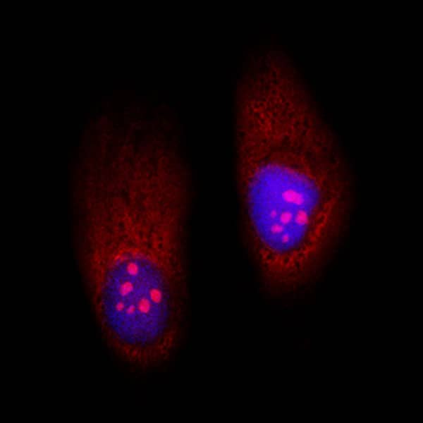
Application of RNA Imaging Probe 1c on PFA-fixed HeLa cells. Confocal fluorescence images of PFA-fixed HeLa cells incubated with 20 μM RNA Imaging Probe 1c (red) and 1 μg/mL Hoechst 33342 (blue) for 30 min.
 View Larger
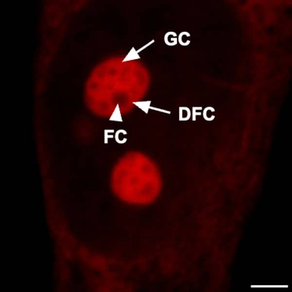
Application of RNA Imaging Probe 1c on HeLa cells. Zoomed-in confocal fluorescence image of HeLa cells stained with 20 μM RNA Imaging Probe 1c for 30 min. Image shows the sub-nucleolar components fibrillar center (FC) (indicated by the white arrowhead), dense fibrillar component (DFC), and outer granular component (GC) (indicated by the white arrows). Scale bar: 3 μm.
 View Larger
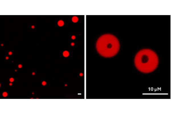
Yeast RNA and spermine labeled with RNA Imaging Probe 1c. Torula yeast RNA and spermine were incubated with RNA Imaging Probe 1c (10 μM). FLIM images show the formation of spherical coacervate droplets upon mixing the RNA and spermine. Image is enlarged by 1× (on the left) or by 8× (on the right). Scale bar: 10 μm.
Sold under license from the University of Southern California.
| λabs | 556 nm |
|---|---|
| λem | 608 nm |
| Extinction Coefficient (ε) | 27500 M-1cm-1 |
| Quantum Yield (φ) | 0.49 |
| Cell Permeable | Yes |
Use our spectra viewer to interactively plan your experiments, assessing multiplexing options. View the excitation and emission spectra for our fluorescent dye range and other commonly used dyes.
Spectral Viewer| M. Wt | 362.21 |
| Formula | C16H15IN2 |
| Storage | Store at -20°C |
| Purity | ≥98% (HPLC) |
| PubChem ID | 171378441 |
| InChI Key | ODMXTRDMWRHIDS-UHFFFAOYSA-M |
| Smiles | C[N+]1=CC=C(/C=C/C2=CC=C3N2C=CC=C3)C=C1.[I-] |
The technical data provided above is for guidance only. For batch specific data refer to the Certificate of Analysis.
Tocris products are intended for laboratory research use only, unless stated otherwise.
| Solvent | Max Conc. mg/mL | Max Conc. mM | |
|---|---|---|---|
| Solubility | |||
| DMSO | 7.24 | 20 |
The following data is based on the product molecular weight 362.21. Batch specific molecular weights may vary from batch to batch due to the degree of hydration, which will affect the solvent volumes required to prepare stock solutions.
| Concentration / Solvent Volume / Mass | 1 mg | 5 mg | 10 mg |
|---|---|---|---|
| 0.2 mM | 13.8 mL | 69.02 mL | 138.04 mL |
| 1 mM | 2.76 mL | 13.8 mL | 27.61 mL |
| 2 mM | 1.38 mL | 6.9 mL | 13.8 mL |
| 10 mM | 0.28 mL | 1.38 mL | 2.76 mL |
References are publications that support the biological activity of the product.
Kim et al (2023) Development of highly fluorogenic styrene probes for visualizing RNA in live cells. ACS Chem.Biol. 18 1523 PMID: 37200527
If you know of a relevant reference for RNA Imaging Probe 1c, please let us know.
Keywords: RNA Imaging Probe 1c, RNA Imaging Probe 1c supplier, RNA, imaging, probe-1c, fluorescent, fluorophore, probe, probes, live, confocal, microscopy, membrane, permeable, fluorogenic, FLIM, photostability, Fluorescent, Probes, 8813, Tocris Bioscience
Citations are publications that use Tocris products.
Currently there are no citations for RNA Imaging Probe 1c. Do you know of a great paper that uses RNA Imaging Probe 1c from Tocris? Please let us know.
There are currently no reviews for this product. Be the first to review RNA Imaging Probe 1c and earn rewards!
$25/€18/£15/$25CAN/¥75 Yuan/¥2500 Yen for a review with an image
$10/€7/£6/$10 CAD/¥70 Yuan/¥1110 Yen for a review without an image
Tocris offers the following scientific literature in this area to showcase our products. We invite you to request* your copy today!
*Please note that Tocris will only send literature to established scientific business / institute addresses.
This product guide provides a background to the use of Fluorescent Dyes and Probes, as well as a comprehensive list of our: