Flow Cytometry
Flow Cytometry (FCM) is a widely used technique for cell counting, fluorescence-activated cell sorting (FACS), cell cycle analysis, identifying cell populations in heterogeneous samples, analyzing cell characteristics and function, as well as measuring cell surface and intracellular molecules. Flow cytometry is an efficient high throughput technique, capable of recording multiple signals at once.
UV or Near UV Laser Line |
|
| Cat. No. | 产品名称/活性 |
|---|---|
| 5748 | DAPI |
| Blue-fluorescent DNA stain | |
| 5117 | Hoechst 33342 |
| Blue-fluorescent dye for DNA staining | |
Violet Laser Line |
|
| Cat. No. | 产品名称/活性 |
| 6489 | Ocean Blue, SE |
| Blue fluorescent dye; supplied as NHS ester | |
Blue Laser Line |
|
| Cat. No. | 产品名称/活性 |
| 7121 | 7-Aminoactinomycin D |
| Red-fluorescent DNA stain | |
| 5119 | Calcein AM |
| Cell permeable compound; hydrolyzed to become fluorescent in living cells | |
| 5935 | H2DCFDA |
| Fluorescent ROS indicator; cell permeable | |
| 5135 | Propidium iodide |
| Red-fluorescent DNA stain; membrane impermeant to live cells | |
Green Laser Line |
|
| Cat. No. | 产品名称/活性 |
| 5440 | FITC |
| Green fluorescent dye | |
| 7313 | Hoechst Janelia Fluor® 526 |
| Fluorogenic, green-emitting DNA probe | |
Yellow Laser Line |
|
| Cat. No. | 产品名称/活性 |
| 6503 | Janelia Fluor® 549, free acid |
| Yellow fluorescent dye; supplied as a free acid | |
| 6500 | Janelia Fluor® 549, Maleimide |
| Cell permeable yellow fluorescent dye; supplied with a maleimide reactive group | |
| 6147 | Janelia Fluor® 549, NHS ester |
| Cell permeable yellow fluorescent dye; supplied as NHS ester | |
| 6502 | Janelia Fluor® 549, Tetrazine |
| Cell permeable yellow fluorescent dye; supplied as a tetrazine for 'click' chemistry | |
| 7693 | MitoBrilliant™ Live 549 |
| Yellow/orange fluorescent mitochondrial stain for live cells, Δψm dependent. | |
Red Laser Line |
|
| Cat. No. | 产品名称/活性 |
| 5436 | Cyanine 5, SE |
| Red fluorescent dye for the labeling of amines; supplied as NHS ester | |
| 6804 | Hoechst Janelia Fluor® 646 |
| Fluorogenic, red-emitting DNA probe | |
| 8027 | Janelia Fluor® 635, Maleimide |
| Red fluorescent dye, supplied with a maleimide reactive group | |
| 7088 | Janelia Fluor® 646, Azide |
| Cell permeable red fluorescent dye; supplied as an azide for 'click' chemistry | |
| 6590 | Janelia Fluor® 646, Maleimide |
| Red fluorescent dye, supplied with a maleimide reactive group | |
| 6148 | Janelia Fluor® 646, NHS ester |
| Cell permeable red fluorescent dye; supplied as NHS ester | |
| 7700 | MitoBrilliant™ 646 |
| Universal red fluorescent mitochondrial stain for both live and fixed cells. | |
| 7417 | MitoBrilliant™ Live 646 |
| Red fluorescent mitochondrial stain for live cells, Δψm dependent. | |
Other |
|
| Cat. No. | 产品名称/活性 |
| 8025 | Janelia Fluor® 525, Maleimide 最新 |
| Cell permeable yellow fluorescent dye; supplied with a maleimide reactive group | |
| 8097 | Janelia Fluor® 669, Maleimide |
| Red fluorescent dye, supplied with a maleimide reactive group | |
| 8915 | Janelia Fluor® 646, free acid 最新 |
| Red fluorescent dye; supplied as a free acid | |
There are several different of types of flow cytometry including spectral flow cytometry, imaging flow cytometry, mass cytometry and cell sorting, but the principles remain the same. Pre-labeled cells are suspended in a buffer and are forced to flow one cell at a time through a flow cytometer instrument. A laser beam is directed at the single cell flow and a detector records the fluorescent and light scatter signals, which are then converted into a plot, allowing researchers to visualize their data.

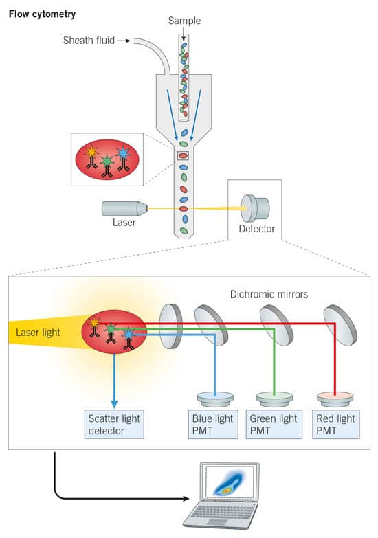
Figure 1: Flow Cytometery Principles: There are three basic flow cytometer components: 1: a fluidics system that uses sheath fluid to force cells through a chamber one cell at a time; 2: an optics system, consisting of a laser that is focused at an interrogation point where the cells will be flowing one at a time. This laser excites the fluorophores conjugated to the labeled antibodies, with the resulting fluorescence emissions passing through a series of dichromic mirrors and optical filters. There are three common optical filters for recording the fluorescence emissions: longpass filters that allow wavelengths over 500 nm through; shortpass filters that allow wavelengths under 600 nm through; and bandpass filters, which allow a specific range of wavelengths through; 3: an electronics system, made up of photo multiplier tubes (PMTs) that capture the fluorescent emission (photons) signal and convert it into an electronic signal. In addition to fluorescence signals from fluorophore-labeled antibodies which identify the presence of a target, a flow cytometer detector also measures forward scatter (FSC) and side scatter (SSC) of the laser light signal. As cells pass in front of the laser, light is scattered, the pattern of this scatter is used to determine the size and nature of cells passing by. FSC is measured in the forward direction (the direction that the laser is traveling) and SSC is measured at a 90° angle. FSC and SSC readings are key controls to remove noise from debris in the cell solution. It is worth noting that the blue laser is usually used to measure light scatter.
Common Flow Cytometry Laser Lines and Selecting Dyes
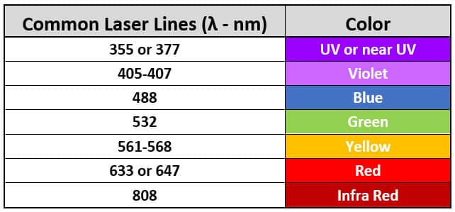
There are several common laser lines used for flow cytometry, and these will govern which dyes are best to use for an experiment. Common laser lines are listed below.
General tips for selecting fluorophores:
- Pick a fluorophore with excitation and emission wavelength close to the laser line of your flow cytometer to ensure maximum efficiency.
- Pick the brightest dye for your lowest-density target and for those that display poor labeling efficiency, particularly when multiplexing.
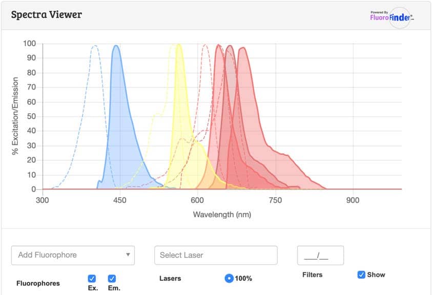
- Ensure minimum spectral contamination "spillover" by selecting dyes with the most distinctly different emission and excitation spectra's possible (use our spectra viewer to plan your experiments).

Figure 2: Common Flow Cytometry Laser Lines.
Flow Cytometry Types
Technological advances in terms of instrument capability and dye properties, have led to new types of flow cytometry applications including:
- Fluorescence Activated Cell Sorting or FACS: As cells pass through a flow cytometer, they are given an electrostatic charge, either positive or negative depending on if the target is detected. Charged cells are then separated, using positively and negatively charged deflection plates, and collected. FACS machines are now able to sort up to 20,000 cells per second.
- Spectral Cytometry: Analyzes the full emission wavelengths of each fluorochrome removing the need for a specific bandpass filter. Algorithms are used to assess proportions of the signal associated with each fluorochrome, which means that the number of dye options is greatly increased, as the researcher is no longer limited by specific bandpass filters.
- Imaging Flow Cytometry: This type of flow cytometry can take an image of the cell at the integration point and measure the fluorescent signal, providing information on the spatial distribution of the target marker in the cell.
- Mass cytometry: Mass cytometry uses antibodies conjugated to stable isotopes with a specific mass that are then measured using time-of-flight (TOF) mass spectrometry. Ideal for use in cell phenotyping panels.
Flow Cytometry Resources
Spectra Viewer
Use our spectra viewer to interactively plan your flow cytometry experiments. View the excitation and emission spectral data for all common flow dyes including Janelia Fluor ® dyes, TFAX (AF) dyes, Cyanine Dyes (Cy Dyes) and BDY (BODIPY®) dyes. Note: absorption spectra may be displayed instead of excitation spectra.

BODIPY® is a registered trademark of Molecular Probes.
Flow Cytometry Panel Builder Tool
Use our sister brands Novus' flow cytometry panel builder tool to design your experiment by enabling researchers to find validated antibodies that work with specific cytometers. Your panel can be exported and saved for reference. Advanced Features of this tool include a spectra viewer, spillover popups and antigen density selector.
Flow Cytometry Handbook from Bio-Techne
Download the Bio-Techne Flow Cytometry eHandbook, full of practical advice, troubleshooting tips and conjugate options.

Complete Flow Cytometry Workflow Solutions
For a comprehensive list of flow cytometry workflow solutions, including flow cytometry-validated pre-conjugated antibodies, Cloudz™ cell selection kits, flow cytometry protocols and Milo™, a single-cell western blotting platform, which can analyze thousands of cells in a single run, visit our sister brand R&D Systems™.
Literature for Flow Cytometry
Tocris offers the following scientific literature for Flow Cytometry to showcase our products. We invite you to request* your copy today!
*Please note that Tocris will only send literature to established scientific business / institute addresses.
Fluorescent Dyes and Probes Research Product GuideUpdated
This product guide provides a background to the use of Fluorescent Dyes and Probes, as well as a comprehensive list of our:
- Fluorescent Dyes, including Janelia Fluor® Dyes
- Fluorescent Probes for Advanced Microscopy and Live Cell Imaging
- Fluorescent Probes to Support In Vivo, Deep Tissue, and Bioluminescence Imaging
- Fluorescent Probes and Reagents for Organoids and 3D Cell Culture Imaging
- Aptamer-based RNA Imaging Reagents
- Fluorescent Probes for Imaging Bacteria
- TSA VividTM Fluorophore Kits
- TSA Reagents for Enhancing IHC, ICC & FISH Signals
- Tissue Clearing Kits and Reagents

