Liver Cancer
Globally, liver cancer is the 2nd leading cause of cancer-related death and the 5th most diagnosed type of cancer. Cancer that originates in the liver can be divided into two types: hepatocellular carcinoma (HCC) accounts for between 80-90% of cases, and cholangiocarcinoma (CCA), which accounts for 10-15% of cases. The liver is also a common site for metastatic secondary cancers that have metastasized from other tumor sites such as the prostate, breast, lung, colon, or pancreas.
Jump to product table
Liver Cancer Product Areas
HCC occurs in hepatocytes and over 90% of HCC cases occur with chronic liver disease. HCC is the leading cause of death in patients with cirrhosis - an advanced form of liver disease. Cirrhosis can occur without any known cause but is more often a result of alcohol consumption, hepatitis infection or non-alcohol related fatty liver disease (NAFLD, also known as steatosis), which can progress to non-alcoholic steatohepatitis (NASH). It is estimated that 2.6% of patients in the US with NASH-associated cirrhosis will develop HCC. When combined with hepatitis B infection, aflatoxin (a naturally occuring toxin produced by some fungi) can also increase the risk of HCC by up to 30 times.
CCA originates in the cholangiocytes of the bile duct. CCA has several associated risk factors, which are largely the same as those for HCC, but it can also occur without any identifiable cause. Specific risk factors for CCA include: infection with liver flukes, cholelithiasis (gallstones) and choledocolithiasis (gallstones in the common bile duct), biliary duct cysts, primary sclerosing cholangitis and hepatitis B and C infection.
Nonalcoholic Fatty Liver Disease
The progression of NAFLD to NASH is a result of liver inflammation and cell death. These processes provide opportunities for liver cancer research, for example, the use of chemokine receptor (CCR) antagonists to reduce hepatic infiltration of inflammatory cytokines. Diabetes often occurs alongside NAFLD, therefore peroxisome proliferator-activated receptors (PPARs), which can be used to target diabetes and are involved in modulating the metabolism of fatty acids and glucose, can also be used in NAFLD research. Other pathways that affect liver cancer development include Wnt and p53 signaling; Wnt activation can speed up progression of liver cancers and p53 is a tumor suppressor gene; loss of function of p53 allows the proliferation and survival of cells that accumulate genetic alterations, leading to tumorigenesis.
Vascular cell adhesion molecules are implicated in the infiltration of inflammatory immune cells into the liver. A significant increase in expression of vascular cell adhesion molecule 1 (VCAM-1) is observed in humans and in mouse models of NASH. VCAM-1 can be used as a biomarker to predict worsening of fibrosis and development of NASH; other cell adhesion molecules have also been shown to correlate with tumor progression.
Glucagon-like peptide 1 (GLP1) receptors are involved in regulating appetite, insulin and glucagon secretion. GLP1 agonists, which are used to treat type 2 diabetes and obesity, modulate the PI3K/AKT/AMPK pathway in mouse models of NAFLD; they may also prove useful in managing NAFLD.
Virus and Parasite Changes in Liver Cancer
Hepatitis B virus (HBV) is estimated to be the cause of around 50% of HCC cases. The use of antiviral drugs has reduced the risk from HBV infection, but further research is required to find effective treatments that target HCC.
Oncogenesis following HBV infection can be triggered by several mechanisms. One pathway results from the production of inflammatory cytokines such as IL-6, TNF-α and TGF-β, which first leads to liver fibrosis, then to cirrhosis and finally to tumorigenesis. Another pathway follows an increase in cell proliferation and evasion of apoptosis, followed by DNA alterations (e.g. p53, MYC proto-oncogene, IDH1, IDH2 and FGFR2) and then either oncogene dysregulation or epigenetic alterations mediated by IDH1 or IDH2. TERT overexpression, an HBV-mediated insertional mutation site, which leads to overexpression of telomerase, preventing senescence and promoting cell transformation is another pathway leading to oncogenesis.
HBV induces infected hepatocytes to produce viral proteins including HBx. HBx has tumorigenic effects including the promotion of cell cycle progression and liver fibrosis, plus inhibition of p53 tumor suppressor. More specifically, HBx upregulates or activates MYC, NF-κB and β-catenin, all of which are involved in promoting carcinogenesis. HBx transgenic mice are more likely to develop HCC than wild-type mice, with some studies showing that 100% of HBx transgenic mice develop HCC.
Infection with liver flukes such as Opisthorchis viverrini is one of the main causes of CCA in parts of the world where infection is common. The fluke worms secrete and excrete several metabolic products, some of which may be toxic or induce inflammatory and immunogenic responses. Mouse fibroblasts show a 4-fold increase in proliferation when co-cultured with adult O. viverrini worms and overexpress mRNA for proteins that induce growth and proliferation, such as TGF. A liver fluke growth factor, granulin, has been shown to activate the MAPK/AKT pathway. Inflammatory cytokines trigger the release of oxygen radicals such as nitric oxide and this can induce DNA damage, inhibit DNA repair, and inhibit apoptosis to promote carcinogenesis.
Tumor Microenvironment Changes in Liver Cancer
Tumors evade normal immune detection by altering the tumor microenvironment (TME) to drive oncogenesis and tumor growth. In liver cancer, the main tumorigenic process is inflammation - driven by alcohol, viral hepatitis and fluke infection. Oncogenic β-catenin signaling is also associated with chronic inflammation.
Research suggests that interferon-γ (IFN-γ) has a dose-dependent effect on tumors: at low levels it is tumorigenic but at higher levels it is anti-tumorigenic, apoptotic and induces autophagy. The combination of lowered IFN-γ levels and reduced infiltration by lymphocytes in liver cancers is associated with worse prognosis. Evidence suggests that serum levels of IFN-γ can also be used as a biomarker for tumor recurrence or severity, with lower IFN-γ levels correlating with higher risk of recurrence and worse outcomes.
The upregulation of immune checkpoints (inhibitory receptors that activate immunosuppressive signaling in immune cells) helps tumor cells to evade immune detection, and this may occur alongside increased expression of indoleamine 2,3 dioxygenase (IDO), which causes localized depletion of amino acids essential for immune cell function.
Genetic and Epigenetic Changes in Liver Cancer
The dominant mutations in HCC are TP53, TERT and CTNNB1. P53/TP53, when inactivated, facilitates tumor progression; TERT is an HBV-mediated insertional mutation site, which leads to overexpression of telomerase, prevents senescence and promotes cell transformation; and CTNNB1 activates Wnt/β-catenin signaling. TP53 and ARID1A mutations are more common in fluke-related CCA; BAPI, IDH1 and IDH2 are more common in non-fluke-related CCA.
Aflatoxin exerts its toxicity through inducing DNA adducts. These cause genetic changes in hepatic cells, DNA strand breakage, DNA base damage and oxidative damage that can lead to tumorigenesis. High exposure to aflatoxin also correlates with HCC due to mutation of p53.
The main epigenetic changes in liver cancer are DNA methylation and histone modification. DNA can be both hypo- and hypermethylated. DNA hypermethylation in fluke-related CCA is primarily at CpG islands; in non-fluke related CCA the hypermethylation occurs at CpG island shores. DNA hypomethylation is the main change in HCC (proliferation class) and promotor hypermethylation is the main epigenetic change in HCC (non-proliferation class). DNA methyltransferase inhibitors are useful tools in cancer research as they can be used to modify these processes.
Histones undergo epigenetic modification through several processes including acetylation, deacetylation, methylation and ubiquitination. These processes are regulated by enzymes including histone deacetylases (HDACs), histone acetyltransferases (HATs), and histone demethylases (KDMs). These epigenetic changes can be targeted in research with compounds that inhibit specific enzymes or protein-protein interactions. For example, HDAC inhibitors, although not effective against tumors when used alone, can be used to sensitize tumor cells to other treatments such as Sorafenib (Cat. No. 6814). Further study on epigenetic changes may uncover other combinations that could be used to treat liver cancers.
Targeted Protein Degradation with PROTAC® Degraders
Most oncogenic cellular, genetic and epigenetic changes are currently undruggable. However, the development of targeted protein degradation provides a way to directly target specific proteins of interest. Proteins that can be targeted for degradation include receptors (both wild-type and mutant), bromodomains, enzymes and other disease-specific proteins. For example, β-catenin can be targeted for degradation with the PROTAC® Degrader xStAx-VHLL (Cat. No. 7298) and can be used in liver cancer research for HBV-initiated liver cancers. A growing range of Degraders is in clinical trials for the potential treatment of a range of cancers and other proteinopathies.
The Role of Lipid Metabolism in Liver Cancer
NAFLD and NASH are associated with significantly increased levels of lipids such as fatty acids, ceramides and triglycerides. One of the animal models of liver cancer is the High-Fat Diet. This model demonstrates steatosis and obesity but not fibrosis, and HCC is rare. Changes in lipid metabolism may be responsible for causing disease, rather than the consumption of a high fat diet. The overexpression of some enzymes and altered levels of some lipids, such as ceramides and sphingolipids, contributes to the generation of a hepatotoxic environment. There is evidence from animal models of NAFLD that the use of statins, such as Rosuvastatin (Cat. No. 6343), reduces HCC development. However, there is also conflicting evidence that low serum cholesterol is associated with an increased risk of HCC.
Models of Liver Cancer in Research
Liver cancer cell lines are one option for screening compounds for potential effectiveness against liver cancer, and animal models of disease provide another way to research compounds in a whole animal system. There are several dietary and non-dietary mouse models for studying NAFLD, NASH and HCC, with the aim of also inducing metabolic syndrome. The 'High-fat, high-cholesterol, high-fructose diet' model, also known as the Western Diet demonstrates steatosis, fibrosis, obesity and HCC development in 89% of the mice, and the diethylnitrosamine plus high-fat model demonstrates nearly 100% disease development. Transgenic mouse models of HBV and HBC infection provide insight into the tumorigenic mechanisms of viral infection and a way to screen potential therapeutic compounds against both tumorigenesis and harmful viral mechanisms.
However, these methods do not fully model the important aspects of human liver biology. Creating organoids from pluripotent stem cells taken directly from a patient more accurately models the tumor structure and environment, and can be used to test treatments that are tailored to a tumor's specific genetics.
New and Top Products for Liver Cancer Research
Click product name to view details and order
| Target | Top Products | New Products |
|---|---|---|
| ATM | KU 55933, VE 821, AZ 5704 | |
| BRAF | SB 590885, Sorafenib, CG 858, SJF 0628 | |
| CTNNB1 | XAV 939 (GMP version available), endo-IWR 1, xStAx-VHLL | WIC1 |
| FGFR | SU 5402, PD 173074, Cediranib, Nintedanib | |
| FLT4 | XL 184 | |
| FXR | GW 4064, GW 3965 | |
| IDH1 | AGI 5198 | |
| KRAS | LC 2, MRTX 849 | AMG 510 |
| MET | SU 5416, Crizotinib, SJF 8240 | |
| PDGFR | Sunitinib, CP 673451, Imatinib, Sorafenib, JNJ 10198409, Cediranib | Linifanib |
| PI3K | LY 294002, Wortmannin, Omipalisib, PI 3065 | |
| RET | SU 5416, SPP 86, Vandetanib, Selpercatinib | |
| VEGFR | Cediranib, Vandetanib | Linifanib |
| WNT | CHIR 99021 (GMP version available), IWP 2, Wnt-C59, LP 922056, WIKI4 (Ancillary Material Grade available) | WIC1, PT-65 |
PROTAC® is a registered trademark of Arvinas Operations, Inc., and is used under license.
Literature for Liver Cancer
Tocris offers the following scientific literature for Liver Cancer to showcase our products. We invite you to request* or download your copy today!
*Please note that Tocris will only send literature to established scientific business / institute addresses.
Cell Cycle and DNA Damage Research Product Guide
This product guide provides a review of the cell cycle and DNA damage research area and lists over 150 products, including research tools for:
- Cell Cycle and Mitosis
- DNA Damage Repair
- Targeted Protein Degradation
- Ubiquitin Proteasome Pathway
- Chemotherapy Targets
Epigenetics Scientific Review
Written by Susanne Müller-Knapp and Peter J. Brown, this review gives an overview of the development of chemical probes for epigenetic targets, as well as the impact of these tool compounds being made available to the scientific community. In addition, their biological effects are also discussed. Epigenetic compounds available from Tocris are listed.
Stem Cells Scientific Review
Written by Kirsty E. Clarke, Victoria B. Christie, Andy Whiting and Stefan A. Przyborski, this review provides an overview of the use of small molecules in the control of stem cell growth and differentiation. Key signaling pathways are highlighted, and the regulation of ES cell self-renewal and somatic cell reprogramming is discussed. Compounds available from Tocris are listed.
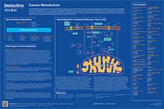
Cancer Metabolism Poster
This poster summarizes the main metabolic pathways in cancer cells and highlights potential targets for cancer therapeutics. Genetic changes and epigenetic modifications in cancer cells alter the regulation of cellular metabolic pathways providing potential cancer therapeutic targets.
Cell Cycle & DNA Damage Repair Poster
In normal cells, each stage of the cell cycle is tightly regulated, however in cancer cells many genes and proteins that are involved in the regulation of the cell cycle are mutated or over expressed. This poster summarizes the stages of the cell cycle and DNA repair. It also highlights strategies for enhancing replicative stress in cancer cells to force mitotic catastrophe and cell death.
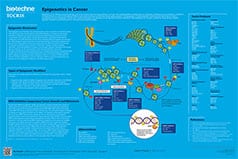
Epigenetics in Cancer Poster
This poster summarizes the main epigenetic targets in cancer. The dysregulation of epigenetic modifications has been shown to result in oncogenesis and cancer progression. Unlike genetic mutations, epigenetic alterations are considered to be reversible and thus make promising therapeutic targets.
Programmed Cell Death Poster
There are two currently recognized forms of programmed cell death: apoptosis and necroptosis. This poster summarizes the signaling pathways involved in apoptosis, necroptosis and cell survival following death receptor activation, and highlights the influence of the molecular switch, cFLIP, on cell fate.
Developing Degraders Poster
This poster describes the generation of a database of Degraders (PROTACs®) from the literature. The Degraders were profiled according to the constituent ligands, linker type, linker length and physicochemical properties and this information was used to establish a set of guidelines for the design and synthesis of cell-permeable Degrader molecules. Presented at the 20th SCI/RSC Medicinal Chemistry Symposium 2019, Cambridge, UK.PROTAC® is a registered trademark of Arvinas Operations, Inc., and is used under license.
Targeted Protein Degradation Poster
Degraders (e.g. PROTACs) are bifunctional small molecules, that harness the Ubiquitin Proteasome System (UPS) to selectively degrade target proteins within cells. They consist of three covalently linked components: an E3 ubiquitin ligase ligand, a linker and a ligand for the target protein of interest. Authored in-house, this poster outlines the generation of a toolbox of building blocks for the development of Degraders. The characteristics and selection of each of these components are discussed. Presented at EFMC 2018, Ljubljana, Slovenia
