Pancreatic Cancer
Pancreatic cancer contributes to 2.5% of all new cancer cases and 4.6% of all cancer deaths worldwide and is one of the most lethal and aggressive cancers, with a 5-year survival rate of less than 9%. Pancreatic cancer is the 4th leading cause of death in developed countries, with these countries also contributing over 50% of new cases. Estimates suggest that pancreatic cancer will become the second major cause of cancer-related deaths. Research is focusing on the tumor microenvironment (TME) and metabolic reprogramming. Early detection of pancreatic cancer is a key area of clinical development, while subtyping, and molecular/genetic landscaping of tumors generates individualized data for integration into treatment modalities to improve survival outcomes.
Jump to product table
Pancreatic Cancer Product Areas
KRAS Mutations and Pancreatic Cancer
Mutations in the Ras GTPase KRAS are activated in over 90% of pancreatic tumors. These mutations and activations are a signature feature of PDAC and act as major drivers of tumor progression, adaptation and therapy resistance. KRAS mutations affect the upregulation of cellular pathways impact the resulting tumor phenotype and clinical outcomes. Pancreatic cancer is one of the most hypoxic cancers and creates a highly immunosuppressive tumor microenvironment (TME), which further increases the challenge of finding effective therapeutic options. The effects of KRAS mutations on cellular mechanisms and on the characteristic traits of PDAC are summarized in Table 1.
Table 1. KRAS mutations associated with pancreatic cancer and their effects on cellular processes, the pathways affected and tools that can target the specific genes or pathways.
| KRAS Mutation (Prevalence) | Trait | Downstream Pathway or Mechanism Affected | Research Tools |
|---|---|---|---|
| G12D (40%) G12V (33%) G12R (15%) Other mutations (12%) | Endocytosis | PI3K-AKT-mTOR RAF-MEK-ERK JNK | Rapamycin - mTOR inhibitor |
| Proliferation | AX 15836 - ERK5 inhibitor | ||
| Invasion | Defactinib - FAK and Pyk2 inhibitor | ||
| Hypoxia | HIF-1α | FM19G11- HIF-1α-subunit inhibitor | |
| Metabolic Changes | Glucose Transporters | BAY 876 - GLUT1 inhibitor | |
| Macropinocytosis | V-ATPase | EN6 - V-ATPase inhibitor | |
| Autophagy | LKB1 > AMPK > ULK1 | MRT 68921 - ULK and autophagy inhibitor | |
| Tumor Microenvironment (immunosuppressive) | TGFβR VEGFR COX2 | SB 431542 - TGFβR inhibitor SU 5416 - VEGFR inhibitor |
Previously considered an 'undruggable target', the direct inhibition of KRAS has been a field of intense interest. The first KRAS PROTAC® LC 2 (Cat. No. 7420) has been developed for targeted protein degradation and creates new opportunities for researching the KRAS-based driving mechanisms of PDAC. Indirect inhibition of KRAS interactions can also be used in research to understand how different pathways contribute to the TME and to PDAC progression. For example, the Ras inhibitor BAY 293 (Cat. No. 6857) and the Raf and MAPK inhibitor Sorafenib (Cat. No. 6814) can indirectly inhibit KRAS interactions.
Tumor Microenvironment and Pancreatic Cancer
Whilst KRAS mutations are important in pancreatic cancer, they should not be considered in isolation but also in combination with the TME and the tumor biology. The hypoxic nature of the TME results in increased HIF1A expression, alterations in cellular metabolism, and local immunosuppression. Inhibition of the hypoxic TME, using compounds such as the HIF-1α inhibitor GN 44028 (Cat. No. 5655), could therefore provide information about immunological changes and reveal new immune-oncology targets. The changes resulting from inhibition of the hypoxic state could also be monitored by using a FAM-labelled HIF-1α peptide (Cat. No. 7452).
The hypoxic nature of the TME leads to a shift in metabolism from oxidative-phosphorylation to glycolysis. The increased glucose requirement associated with this is frequently associated with increased glucose transporter expression; increased expression of GLUT1 is also associated with overexpression of RAS and BRAF genes. The effect of inhibited glucose metabolism on glucose uptake can be monitored through inhibition of GLUT1 using BAY 876 (Cat. No. 6199) in combination with the fluorescent glucose uptake indicator 2-NBDG (Cat. No. 6065). Such approaches could also give insights into tumor metabolic adaptation. Expression changes and tumor evolution or resistance mechanisms could be further evaluated using more broad inhibition of KRAS with Salirasib (Cat. No. 4989) and VEGFR using Axitinib (Cat. No. 4350), whilst monitoring expression levels using ISH, forming a well-integrated workflow.
The SUMO pathway is also an important pathway in PDAC, especially in connection with MYC expression which is a known cancer driver. The SUMOylation of proteins can function as a protective role in hypoxia and other stress states. By modulating SUMO with activators, such as N106 (Cat. No. 5681), or inhibitors like HODHBt (Cat. No. 6994) the effect of SUMOylation on the hypoxic response can be studied further.
Pancreatic Tumor Organoids
Another option for studying pancreatic cancer is to take cells from either healthy pancreas or from a pancreatic tumor and culture these in the presence of a synthetic hydrogel scaffold to produce miniature 3-dimensional organs, or pancreatic organoids. Cells taken directly from a patient with a specific type of tumor and cultured to produce a patient-derived organoid could provide a more direct way to assess how well an individual tumor will respond to treatment. Organoids grown in this way can mimic how tumors would grow and invade surrounding tissues so it may be possible to study how the tumor binds to other tissues and metastasises. By altering the structural supports, for example by inhibiting adhesion, it may be possible to explore how a tumor responds to various treatments.
Related Resources from our Sister Brands
Defining the KRAS mutational status is a challenge using traditional immunohistochemistry approaches, so the use of additional technologies such as BaseScope™ can be used to visualise the mutational state within cells or tissues, and to locate pathways or mutations of interest. Another integrated approach involves the use of liquid biopsies to combine exosome enrichment and BaseScope™ to define the mutational state of a sample. For example, the detection and localization of mutations such as G12V (BA-Hs-KRAS-G12V) and G12C (BA-Hs-KRAS-G12C) could be compared. Another example would be to target the activated PI3K pathway using the PI3K inhibitor Omipalisib (Cat. No. 6792) in the presence of the G12D mutation. RNA/BaseScope™ could also be used to spatio-temporally track tumor evolution, understand the response to treatment and to identify differentially expressed genes and targets of interest.
PROTAC® is a registered trademark of Arvinas Operations, Inc., and is used under license.
New and Top Products for Pancreatic Cancer Research
Click product name to view details and order
| Target | Top Products | New Products |
|---|---|---|
| KRAS | LC 2, BAY 293 | MRTX 849 |
| EGFR | Erlotinib | EMI 48 |
| AKT | AT 7867 | |
| VTPase | SB 590885 | |
| SMARCA4, SMARCA2 | SGC SMARCA-BRDVIII | FHT 1015, FHT 2344 |
| ERBB4 | Neratinib | |
| STAT3 | A 419259 | |
| JAK1 | PKF 115584, FH 535 | |
| MYC | KJ Pyr 9 | |
| mTOR | Torin1, Torin 2 | |
| NOTCH | DBZ | |
| PTEN | SL 327 | |
| Wee1 | Adavosertib |
Literature for Pancreatic Cancer
Tocris offers the following scientific literature for Pancreatic Cancer to showcase our products. We invite you to request* or download your copy today!
*Please note that Tocris will only send literature to established scientific business / institute addresses.
Cell Cycle and DNA Damage Research Product Guide
This product guide provides a review of the cell cycle and DNA damage research area and lists over 150 products, including research tools for:
- Cell Cycle and Mitosis
- DNA Damage Repair
- Targeted Protein Degradation
- Ubiquitin Proteasome Pathway
- Chemotherapy Targets
TPD and Induced Proximity Research Product Guide
This brochure highlights the tools and services available from Bio-Techne to support your Targeted Protein Degradation and Induced Proximity research, including:
- Active Degraders
- TAG Degradation Platform
- Degrader Building Blocks
- Assays for Protein Degradation
- Induced Proximity Tools
RAS Oncoproteins Scientific Review
Written by Kirsten L. Bryant, Adrienne D. Cox and Channing J. Der, this review provides a comprehensive overview of RAS protein function and RAS mutations in cancer. Key signaling pathways are highlighted and therapeutic vulnerabilities are explored. This review also includes a detailed section on RAS drug discovery and targeting synthetic lethal interactors of mutant RAS. Compounds available from Tocris are listed.
Stem Cells Scientific Review
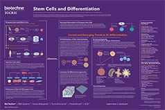
Written by Kirsty E. Clarke, Victoria B. Christie, Andy Whiting and Stefan A. Przyborski, this review provides an overview of the use of small molecules in the control of stem cell growth and differentiation. Key signaling pathways are highlighted, and the regulation of ES cell self-renewal and somatic cell reprogramming is discussed. Compounds available from Tocris are listed.
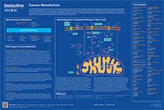
Cancer Metabolism Poster
This poster summarizes the main metabolic pathways in cancer cells and highlights potential targets for cancer therapeutics. Genetic changes and epigenetic modifications in cancer cells alter the regulation of cellular metabolic pathways providing potential cancer therapeutic targets.
Cell Cycle & DNA Damage Repair Poster
In normal cells, each stage of the cell cycle is tightly regulated, however in cancer cells many genes and proteins that are involved in the regulation of the cell cycle are mutated or over expressed. This poster summarizes the stages of the cell cycle and DNA repair. It also highlights strategies for enhancing replicative stress in cancer cells to force mitotic catastrophe and cell death.
Epigenetics in Cancer Poster
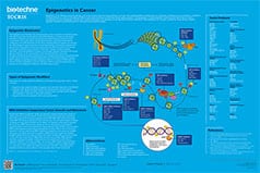
This poster summarizes the main epigenetic targets in cancer. The dysregulation of epigenetic modifications has been shown to result in oncogenesis and cancer progression. Unlike genetic mutations, epigenetic alterations are considered to be reversible and thus make promising therapeutic targets.
Stem Cell Workflow Poster
Stem cells have potential as a source of cells and tissues for research and treatment of disease. This poster summarizes some key protocols demonstrating the use of small molecules across the stem cell workflow, from reprogramming, through self-renewal, storage and differentiation to verification. Advantages of using small molecules are also highlighted.
